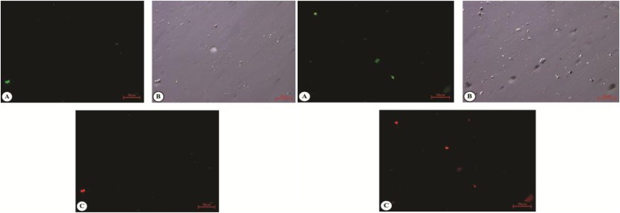
Fig. 10. Images of TSP1KO (Left) and Wild luteal (Right) cells showing signal for FITC Annexin (A) and PI (C). Both the group of cells was cultured for 72 hour followed by trypsinization and treatment with Annexin V apoptosis detection and Propidium iodide dye.After incubation Fluorescent signals were detected by Axio Observer.Z1 microscope. Image (B) indicates cells under bright field image.